This page contains a continually-updated list of findings from the Forensic Metascience AI toolkit's image analysis system which we have submitted to PubPeer. Every finding undergoes thorough human review by Dan Elton and Greg Fitzgerald before submission.
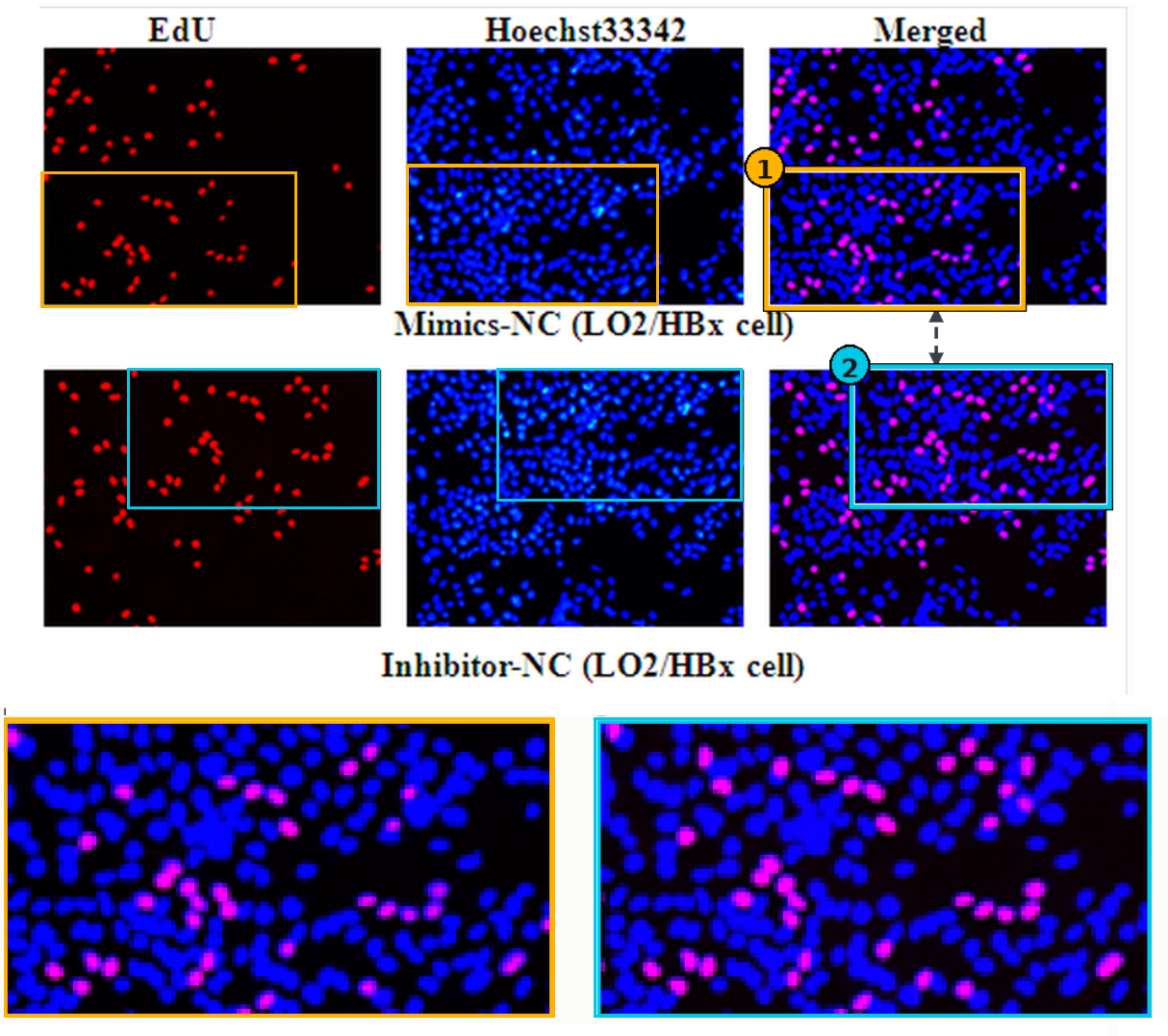
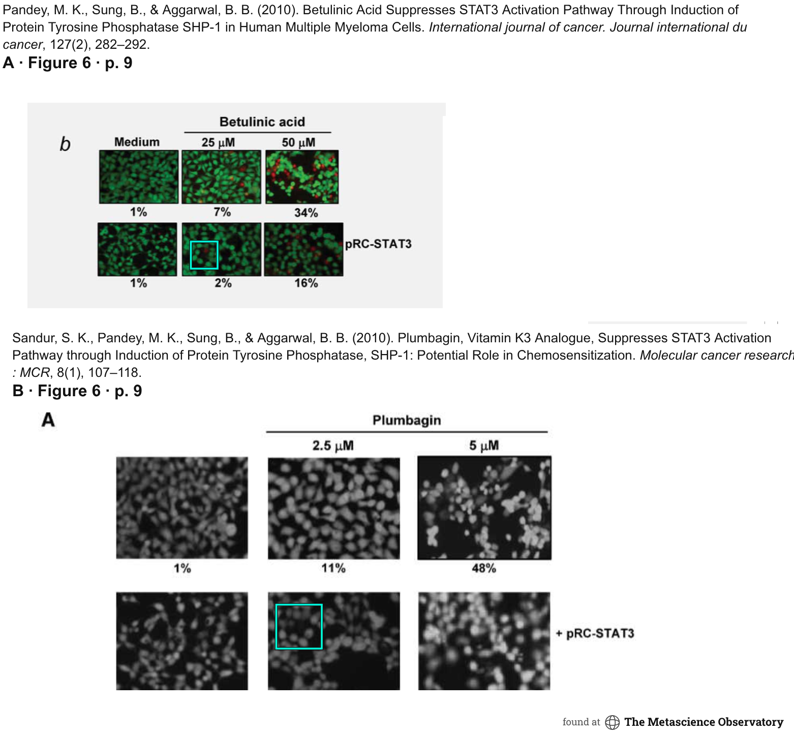
Issue submitted: Figure 5a contains a duplicated region across rows labeled as different treatments. The center column shows matching cells, with almost identical fluorescence patterns, although some spots in the second row do not appear in the first.
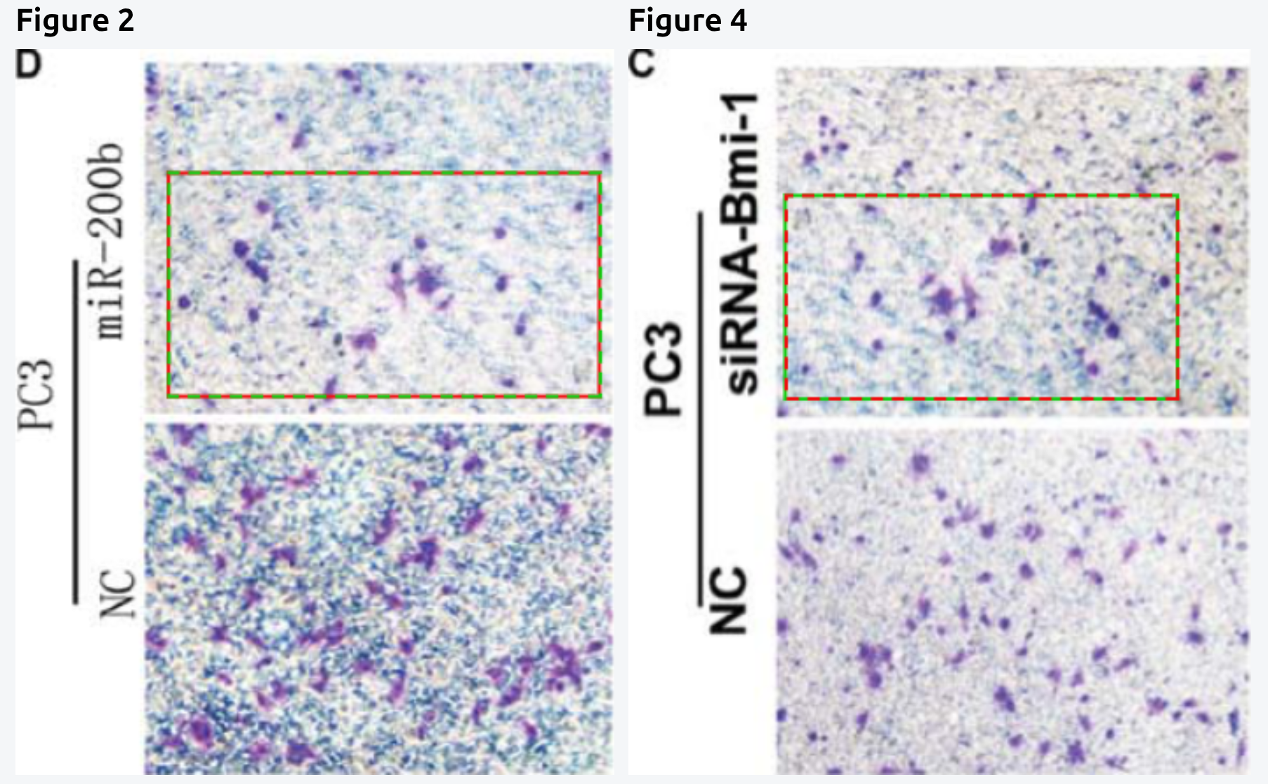
Stephan P. Tenbaum; Paloma Ordóñez-Morán; Isabel Puig; Irene Chicote; Oriol Arqués; Stefania Landolfi; Yolanda Fernández; José Raúl Herance; Juan D. Gispert; Leire Mendizabal; Susana Aguilar; Santiago Ramón y. Cajal; Simó Schwartz; Ana Vivancos; Eloy Espín; Santiago Rojas; José Baselga; Josep Tabernero; Alberto Muñoz; Héctor G. Palmer. Nature Medicine18(6): 892–901 (2012). doi:10.1038/nm.2772
Issue submitted: The cells in Figure 1 are described as coming from a human patient, while the matching cells in Figure 6 are described as coming from a tumor grown subcutaneously in a mouse from human cells.
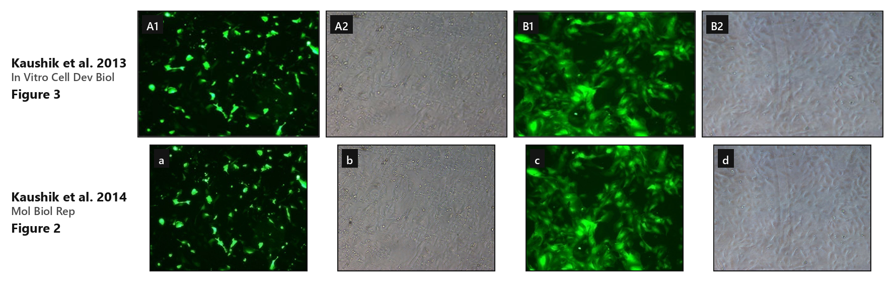
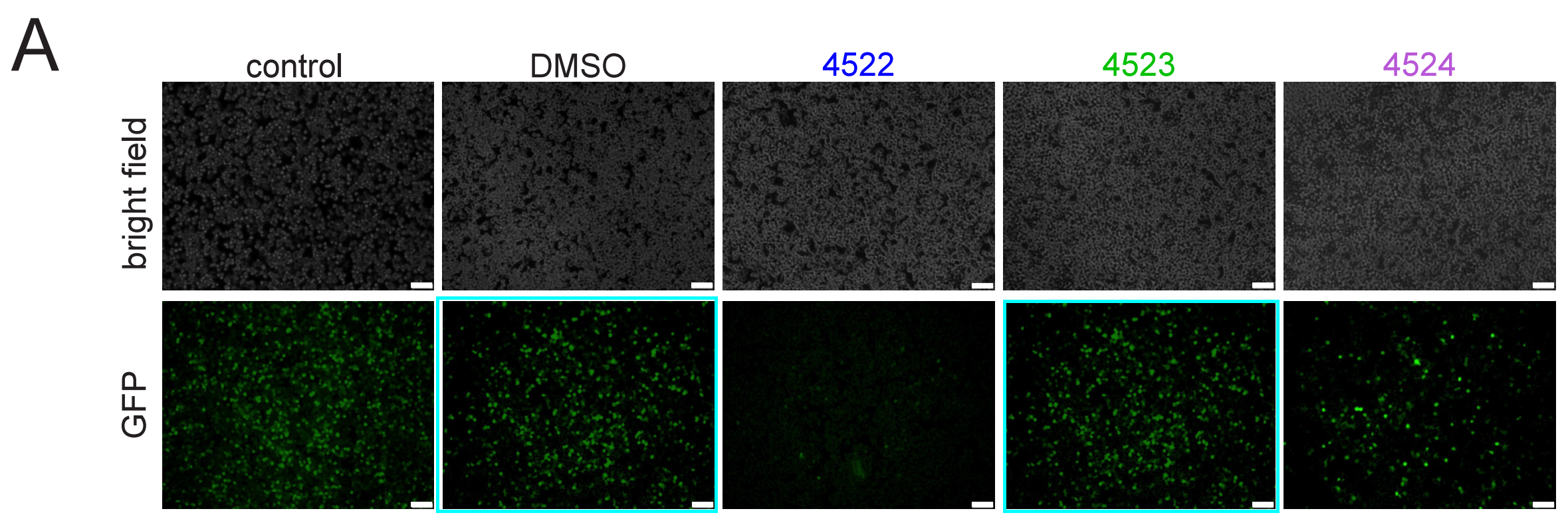
Issue submitted: Images appear to be reused across papers. The 2014 caption describes “EGFP expression in pAcISUBC transfected BuMECs.” The pAcISUBC human insulin expression vector was constructed in the 2014 paper and does not appear in the 2013 paper.
Cara D. Kirby; Gai Liu; Farheen Fatma; Maris R. Pedlow; Narendran G-Dayanandan; Nitin Sharma; Zachary D. Frey; Rachael E. Rush; Steven C. Cardinale; Andrzej M. Krezel; Amy L. Hartman; Terry L. Bowlin; Daisy W. Leung; Gaya K. Amarasinghe. ACS Infectious Diseases11(9): 2373–2382 (2025). doi:10.1021/acsinfecdis.5c00088
Travis L. Stiles; Travis L. Dickendesher; Alban Gaultier; Anthony Fernandez-Castaneda; Elisabetta Mantuano; Roman J. Giger; Steven L. Gonias. Journal of Cell Science126(1): 209–220 (2013). doi:10.1242/jcs.113191
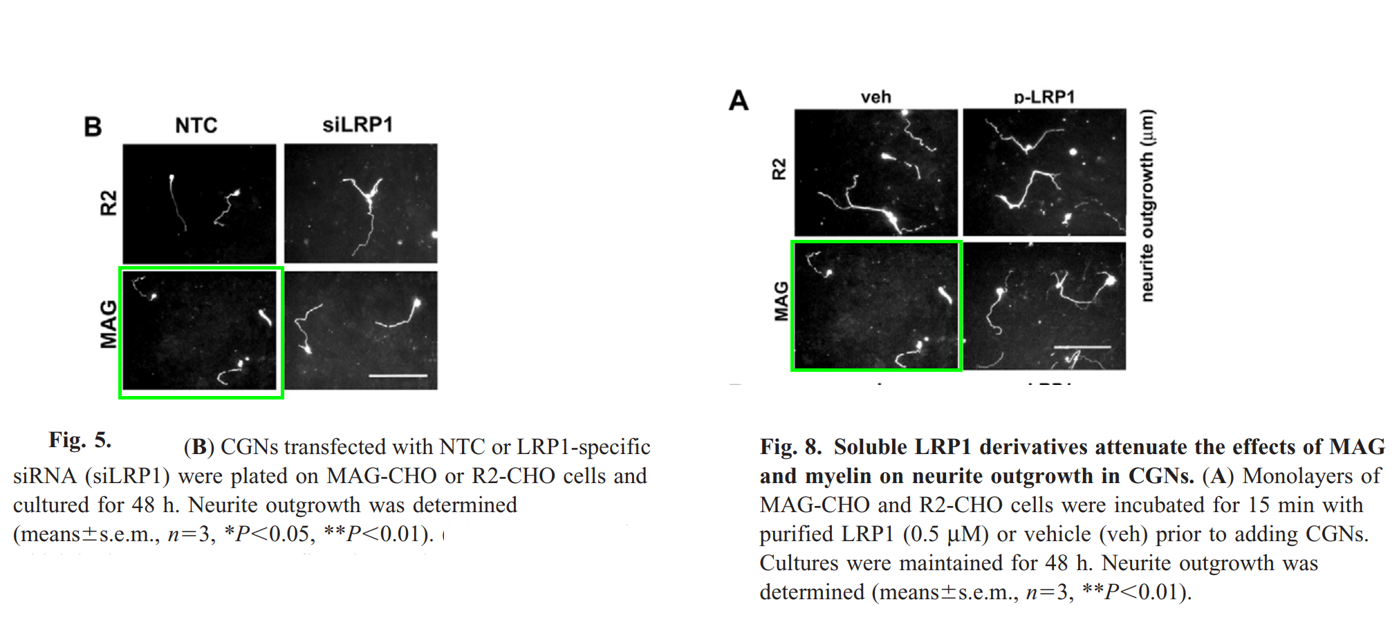
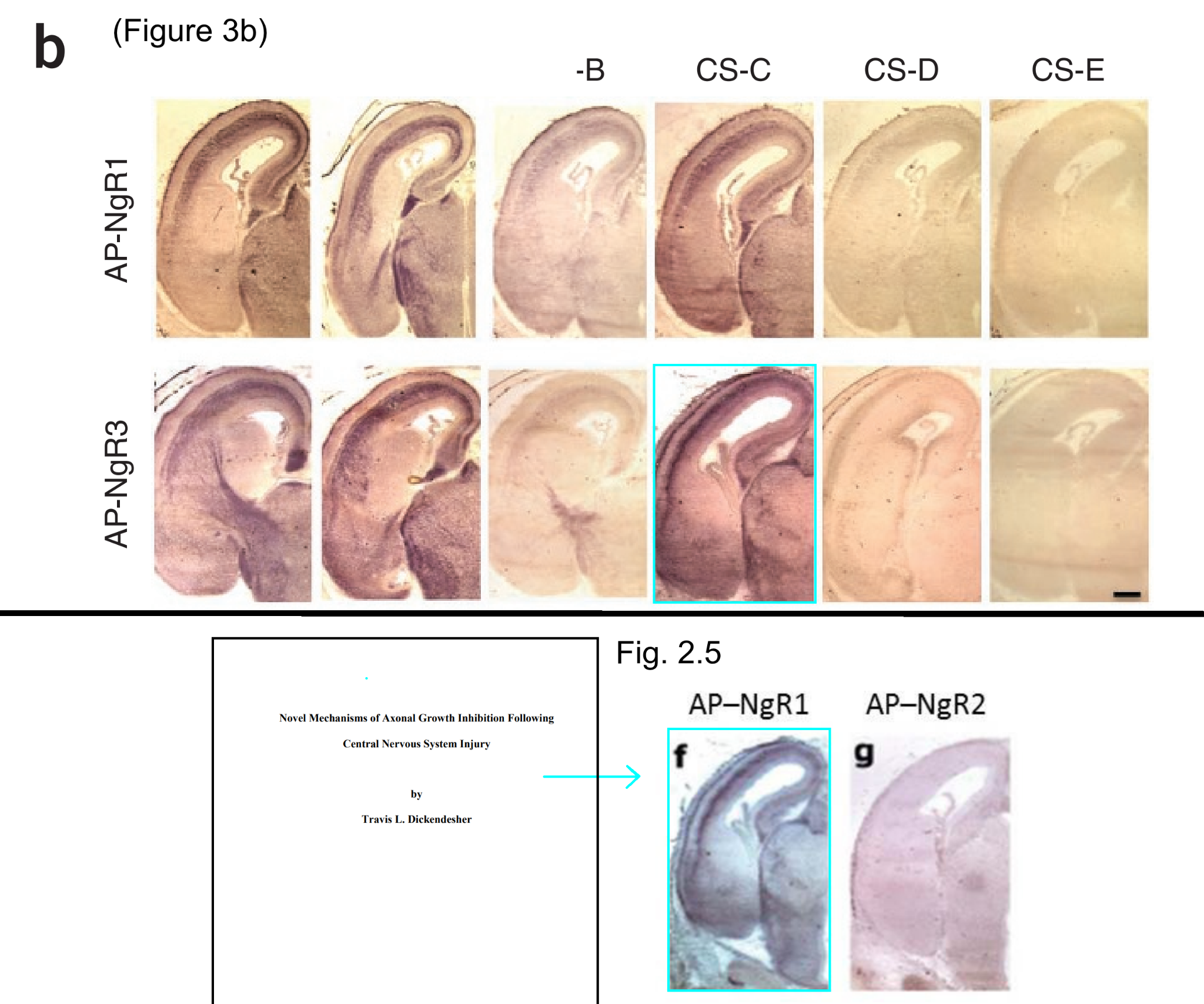
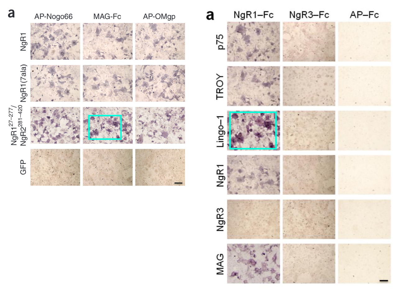
Issue submitted: A duplicated region appears across two separate experiments: the MAG/NTC panel in Figure 5B matches the MAG/vehicle panel in Figure 8A. A panel in Figure 3b also appears in Travis L. Dickendesher’s PhD thesis with a different label and different coloration.
Submitted .
Figures 5B and 8AFigure 3b and Dickendesher thesis Fig. 2.5
Travis L. Dickendesher; Katherine T. Baldwin; Yevgeniya A. Mironova; Yoshiki Koriyama; Stephen J. Raiker; Kim L. Askew; Andrew Wood; Cédric G. Geoffroy; Binhai Zheng; Claire D. Liepmann; Yasuhiro Katagiri; Larry I. Benowitz; Herbert M. Geller; Roman J. Giger. Nature Neuroscience15(5): 703–712 (2012). doi:10.1038/nn.3070
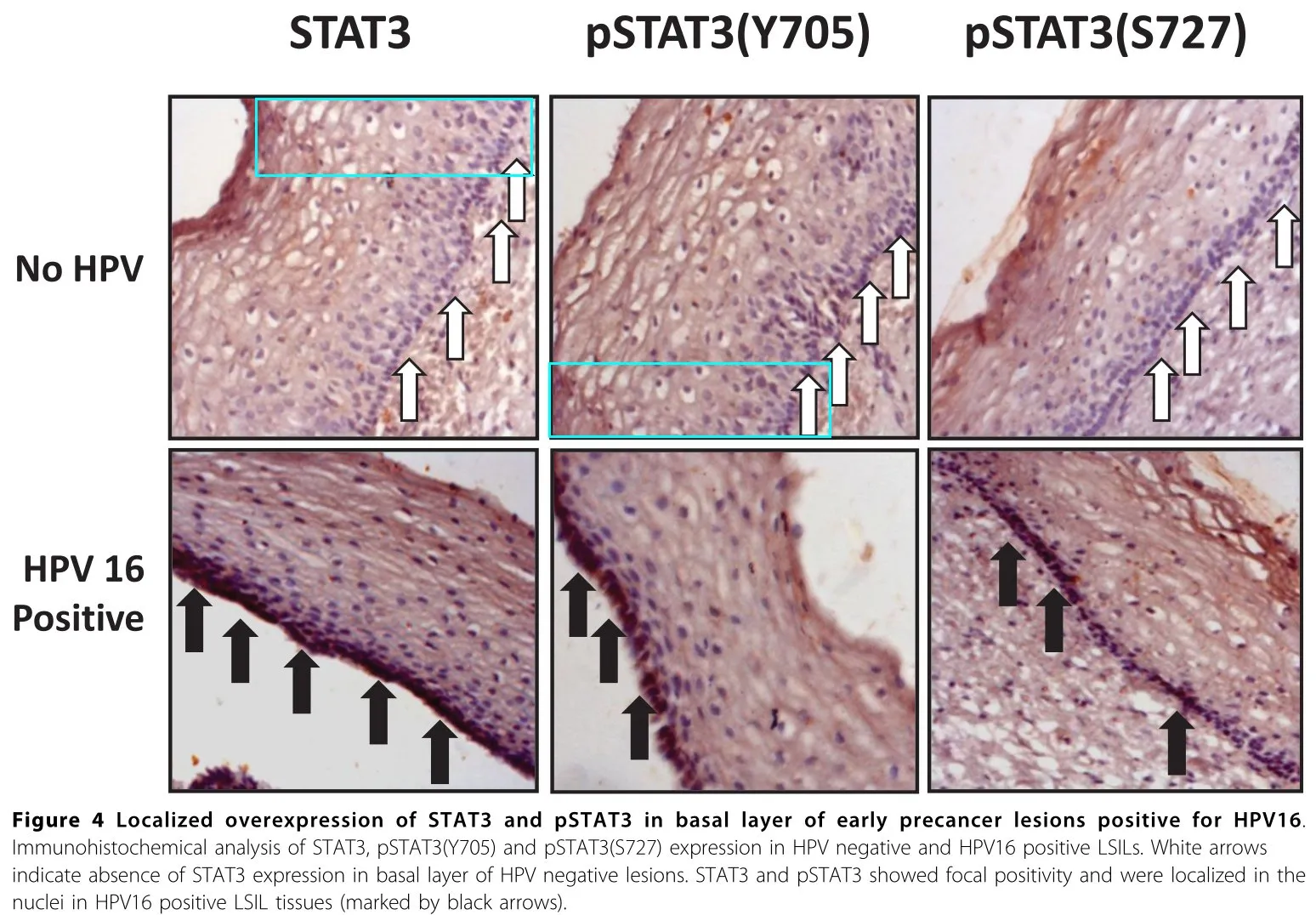
Issue submitted: A duplicated region appears between Supplementary Figure 4 (left) and Figure 4 (right), in addition to issues already noted on PubPeer.
Bryce W. Duncan; Vishwa Mohan; Sarah D. Wade; Young Truong; Alexander Kampov-Polevoi; Brenda R. Temple; Patricia F. Maness. Molecular Neurobiology58(8): 3817–3834 (2021). doi:10.1007/s12035-021-02373-2
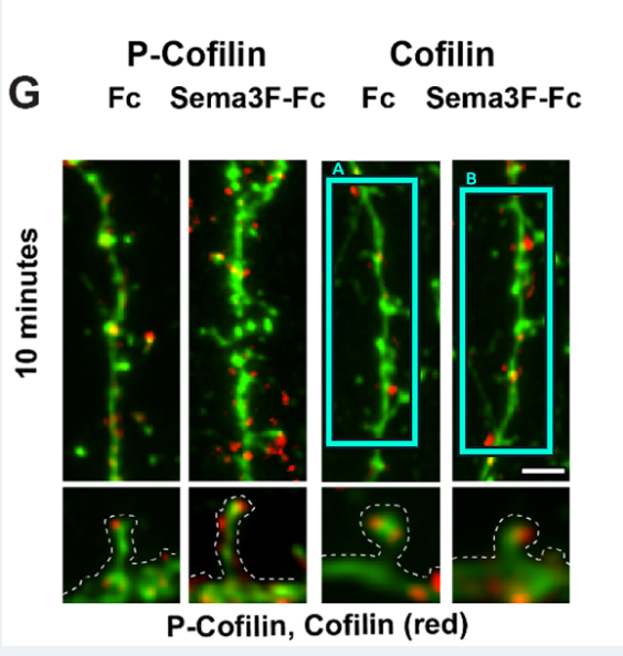
Issue submitted: In figure 3G the panel on the right has the same neurons as the panel next to it, but rotated 180 degrees and then flipped horizontally. The red overlay is similar but a bit different.
Sebum Lee; Yulei Shang; Stephanie A. Redmond; Anatoly Urisman; Amy A. Tang; Kathy H. Li; Alma L. Burlingame; Ryan A. Pak; Ana Jovičić; Aaron D. Gitler; Jinhua Wang; Nathanael S. Gray; William W. Seeley; Teepu Siddique; Eileen H. Bigio; Virginia M.-Y. Lee; John Q. Trojanowski; Jonah R. Chan; Eric J. Huang. Neuron91(1): 41–55 (2016). doi:10.1016/j.neuron.2016.05.021
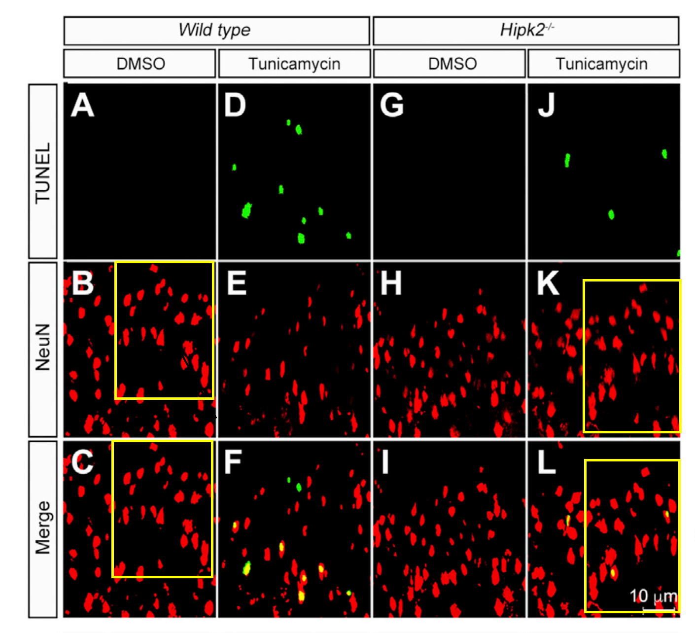
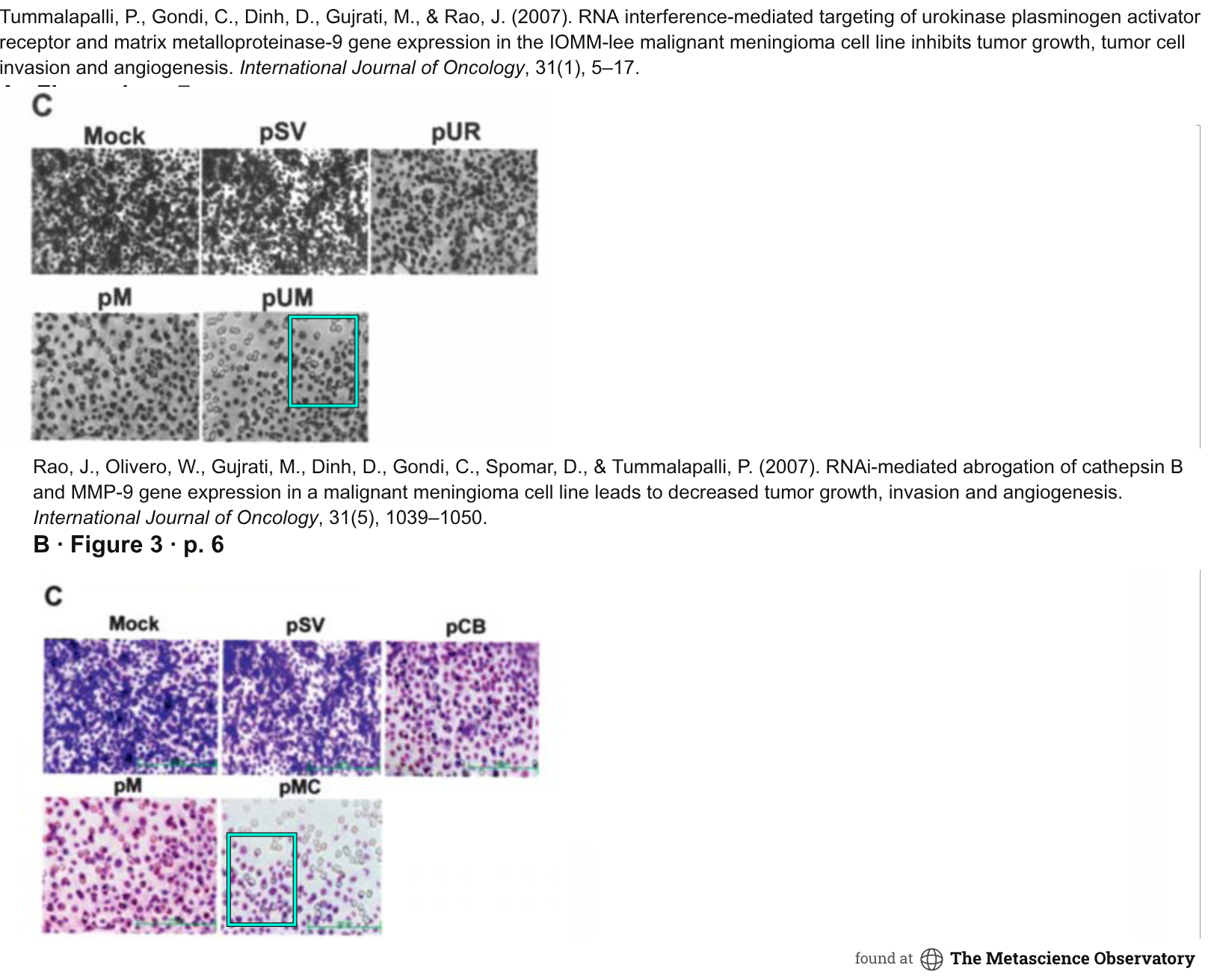
Issue submitted: Figure 1 contains a duplication across conditions, with the image flipped horizontally. Matching regions appear in the wild-type DMSO and Hipk2 knockout tunicamycin panels.
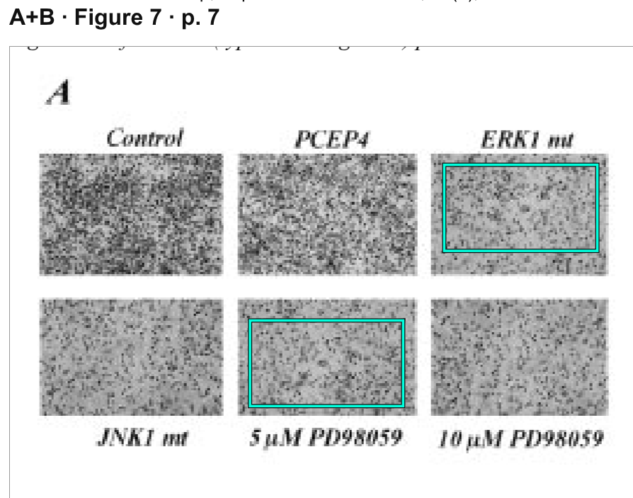
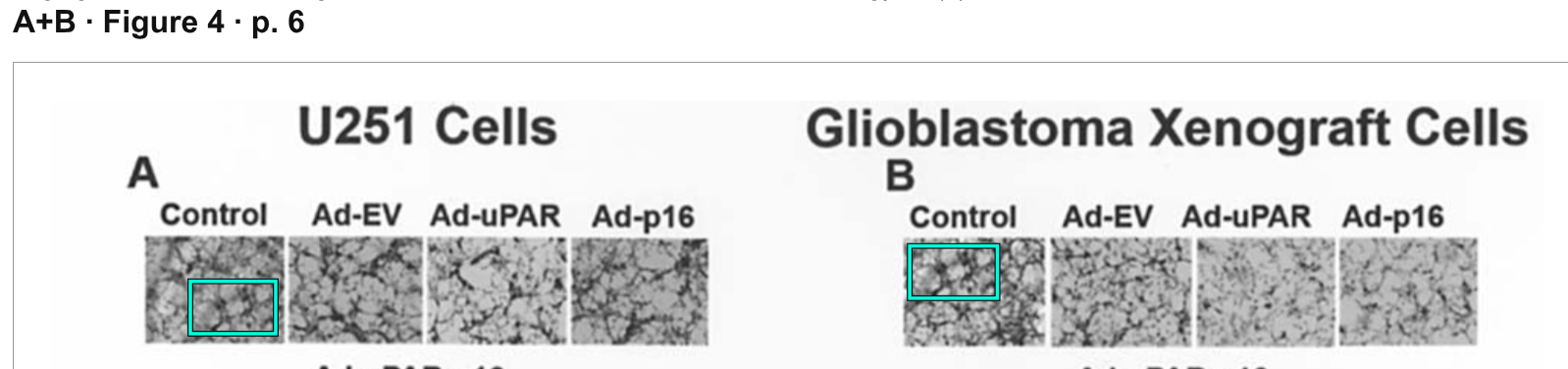
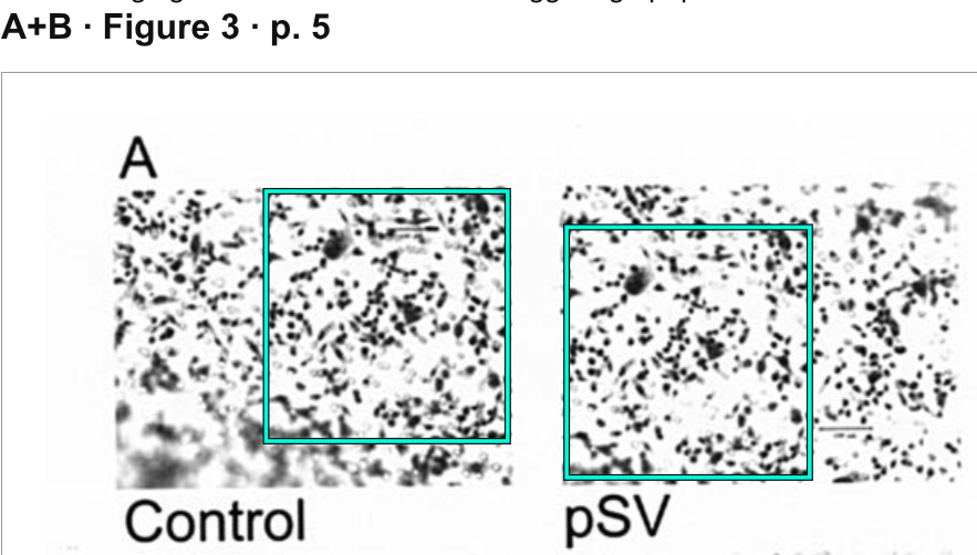
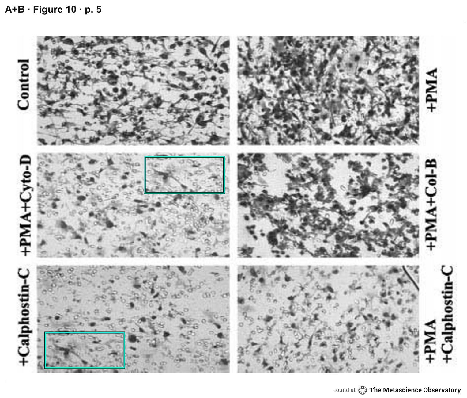
Sajani S. Lakka; Sushma L. Jasti; Anthanassios P. Kyritsis; W.K. Alfred Yung; Francis Ali-Osman; Garth L. Nicolson; Jasti S. Rao. Clinical & Experimental Metastasis18(3): 245–252 (2000). doi:10.1023/a:1006724826083
Ramesh Subramanian; Christopher Gondi; Sajani Lakka; Aman Jutla; Jasti Rao. International Journal of Oncology28(4): 831–839 (2006). doi:10.3892/ijo.28.4.831
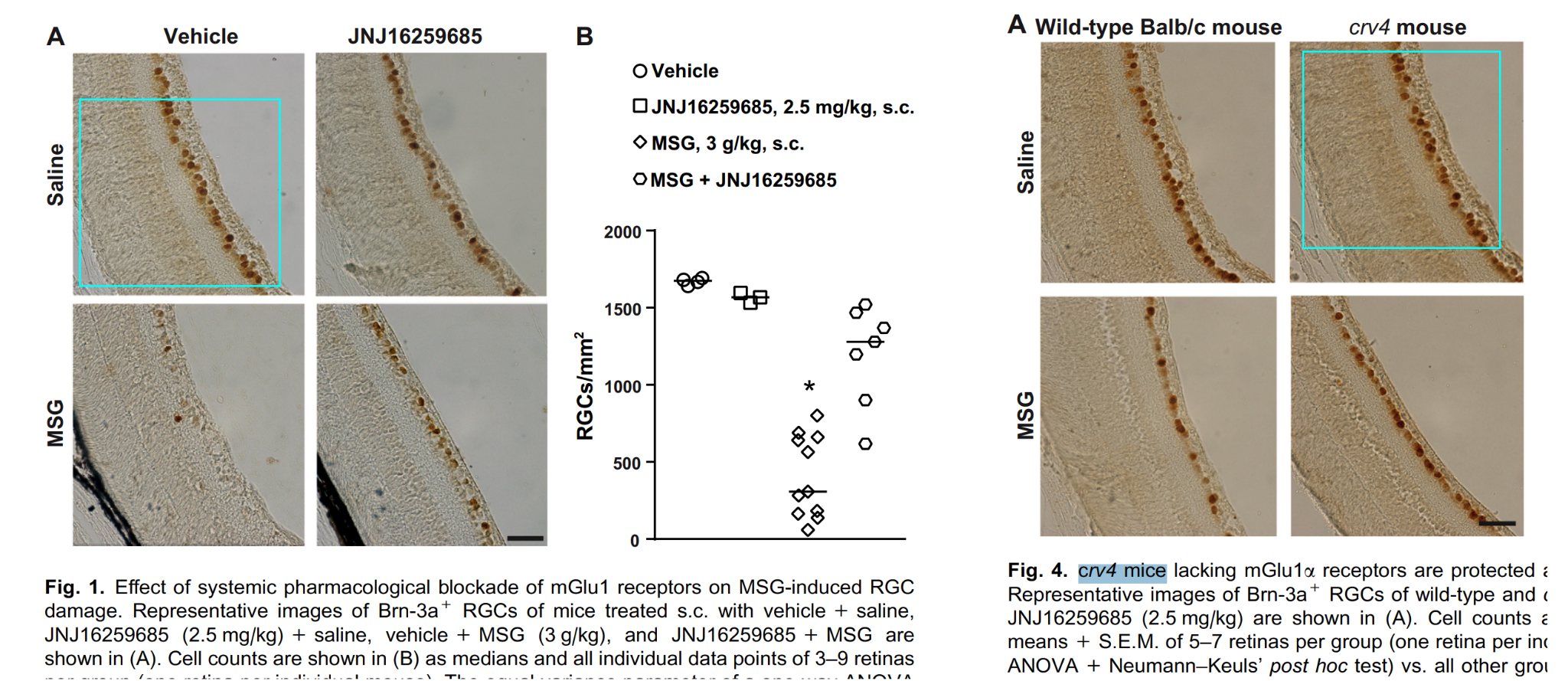
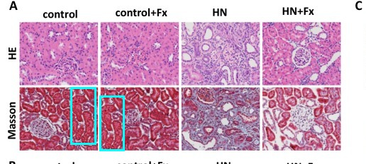
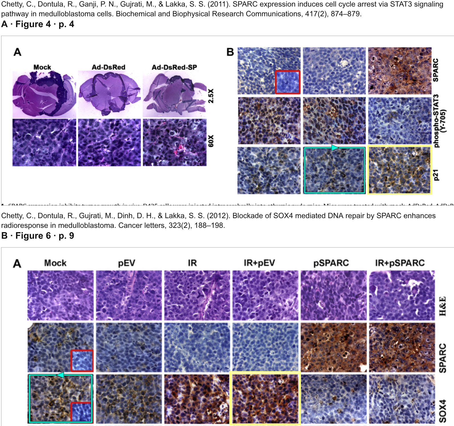
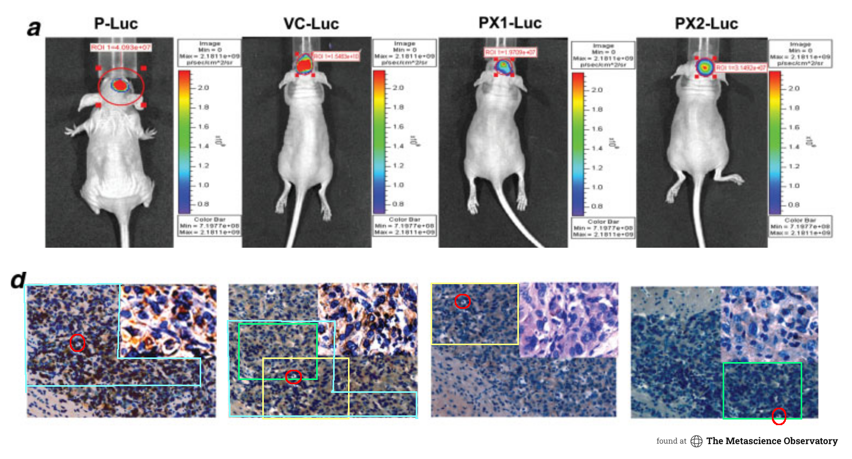
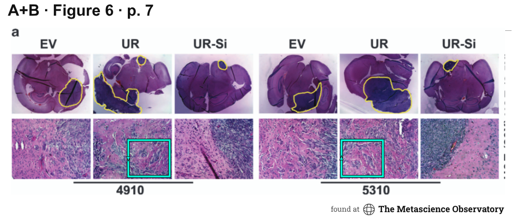
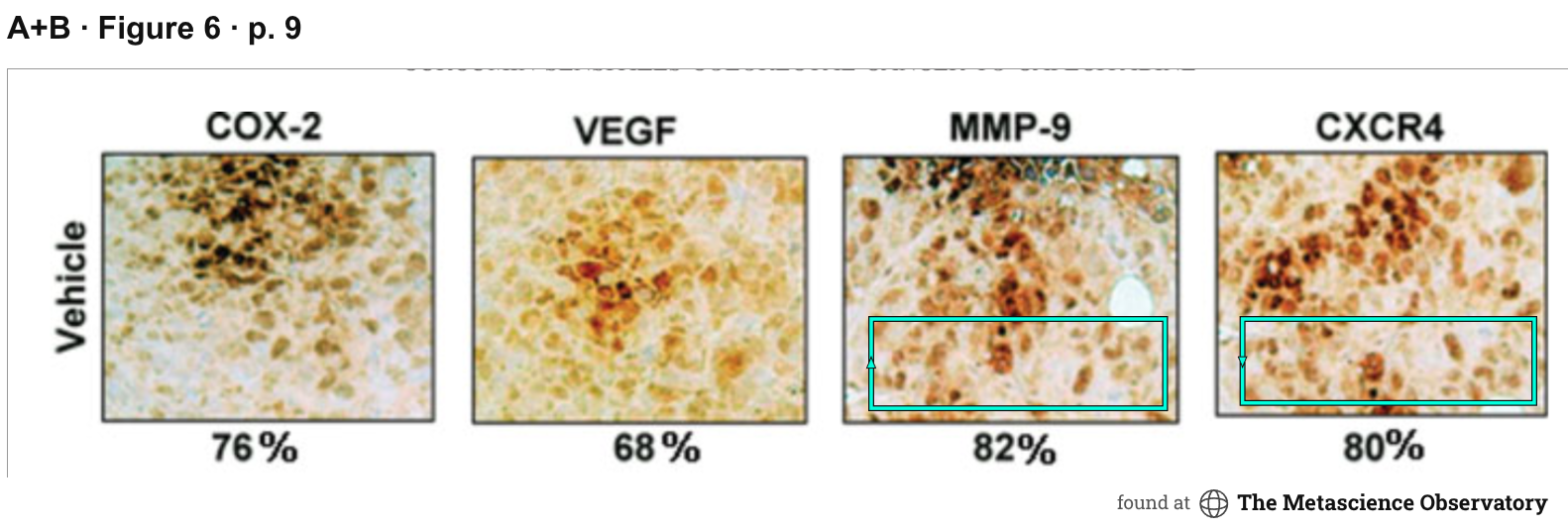
Issue submitted: An image is re-used from another publication with a 90 degree rotation. The image purports to show xenographed tumor tissue across four different mice, but the same tissue appears in all four images but with changes in color.
Submitted .
Earlier comparison: image reuse across publications with a 90-degree rotation. Submitted September 26, 2026.Tumor tissue comparison across four mice. Submitted September 27, 2026.
Ajaikumar B. Kunnumakkara; Parmeswaran Diagaradjane; Preetha Anand; Harikumar B. Kuzhuvelil; Amit Deorukhkar; Juri Gelovani; Sushovan Guha; Sunil Krishnan; Bharat B. Aggarwal. International Journal of Cancer125(9): 2187–2197 (2009). doi:10.1002/ijc.24593
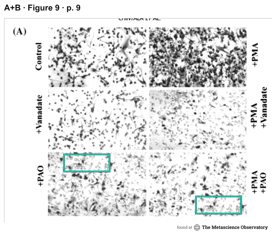
Shravan K. Chintala; Athanassios P. Kyritsis; Pamarthi M. Mohan; Sanjeeva Mohanam; Raymond Sawaya; Ziya Gokslan; W. K. Alfred Yung; Peter Steck; Joon H. Uhm; Bharat B. Aggarwal; Jasti S. Rao. Molecular Carcinogenesis26(4): 274–285 (1999). doi:10.1002/(sici)1098-2744(199912)26:4<274::aid-mc6>3.0.co;2-1
Shravan K. Chintala; Raymond Sawaya; Bharat B. Aggarwal; Sadhan Majumder; Dipak K. Giri; Athanassios P. Kyritsis; Ziya L. Gokaslan; Jasti S. Rao. Journal of Biological Chemistry273(22): 13545–13551 (1998). doi:10.1074/jbc.273.22.13545